

New findings on brain stiffness in multiple sclerosis - Measuring tissue rigidity could improve MS monitoring
What was the research question or scientific inquiry behind your study?
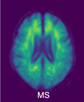
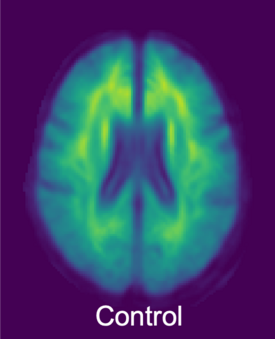
Magnetic resonance imaging (MRI) is routinely used to diagnose and monitor multiple sclerosis (MS). However, MRI cannot easily detect changes in the brain’s gray matter, which is known to contribute to the development of MS symptoms. Our aim was to improve detection of gray matter damage by using tomoelastography, a novel imaging technique developed at Charité that can detect changes in tissue stiffness. We already knew from our previous work that MS leads to changes in brain stiffness. In this study, we investigated whether stiffness changes, particularly in the gray matter, can be used to detect disease activity.
How did you approach the topic?
Tomoelastography is an MRI-based noninvasive technique developed in our group that can be used to measure brain stiffness in both humans and mice. In the field of medical imaging, this is an extremely valuable property because it allows us to map brain stiffness at the micrometer scale in small animals in preclinical research and at coarser resolution in clinical MRI scanners in patients. We used this unique capability to decipher the mechanical changes in the mouse brain and thus learn more about the processes in humans.
What did you discover?
We show that the cortex, i.e. the outer layer of the brain that is made up of gray matter, is softer in MS patients than in healthy people. In the animal model of MS, we discovered that changes to the perineuronal nets are responsible for the softening. Perineuronal nets are a mesh that surrounds neurons and becomes loosened during inflammation, thereby affecting the connections between neurons and possibly causing cognitive problems. The good news is that in the animal model softening seems to be reversible; once inflammation ceases, the perineuronal nets and the stiffness recover.
Was there anything that surprised you?
Yes, we were surprised to see the consistency of the effect of cortical softening across species. The same effect we identified in the mouse model was present in humans.
What’s your takeaway?
This study suggests that tomoelastography could be used to detect the cortical changes that occur in MS and perhaps to determine whether treatments are effective in individual patients. The study also highlights the importance of studying gray matter changes in MS, as changes in the cortex correlate with cognitive symptoms and early disease manifestations. Using tomoelastography to detect changes in mechanical properties in the brain provides a new way to monitor disease activity and therapeutic drug efficacy in MS.
Source
Silva RV et al. Cortical matrix remodeling as a hallmark of relapsing-remitting neuroinflammation in MR elastography and quantitative MRI. Acta Neuropathol 2024 Jan 4. doi:10.1007/s00401-023-02658-x
Contact
Prof. Carmen Infante Duarte
Experimental and Clinical Research Center
Charité – Universitätsmedizin Berlin
Prof. Ingolf Sack
Department of Radiology
Charité – Universitätsmedizin Berlin
Links
- Link to the original article
- Link to the MDC website of the Infante Duarte Lab (Innate Immunity & Neuroinflammation)
- Link to the website of the Sack Lab (Elastography)
